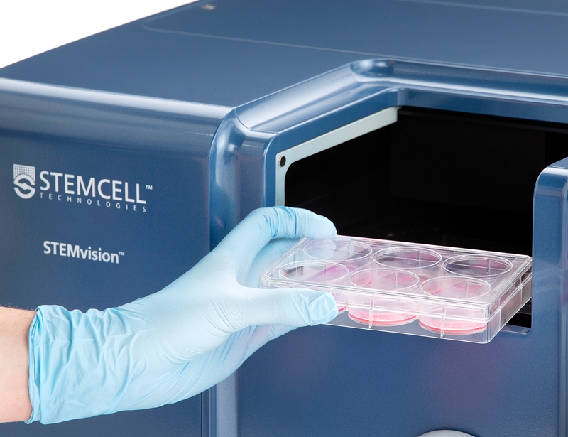
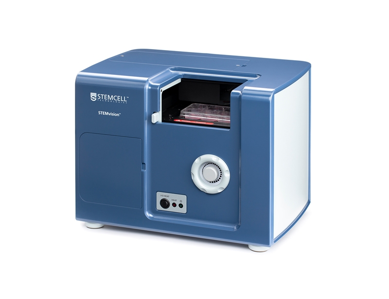
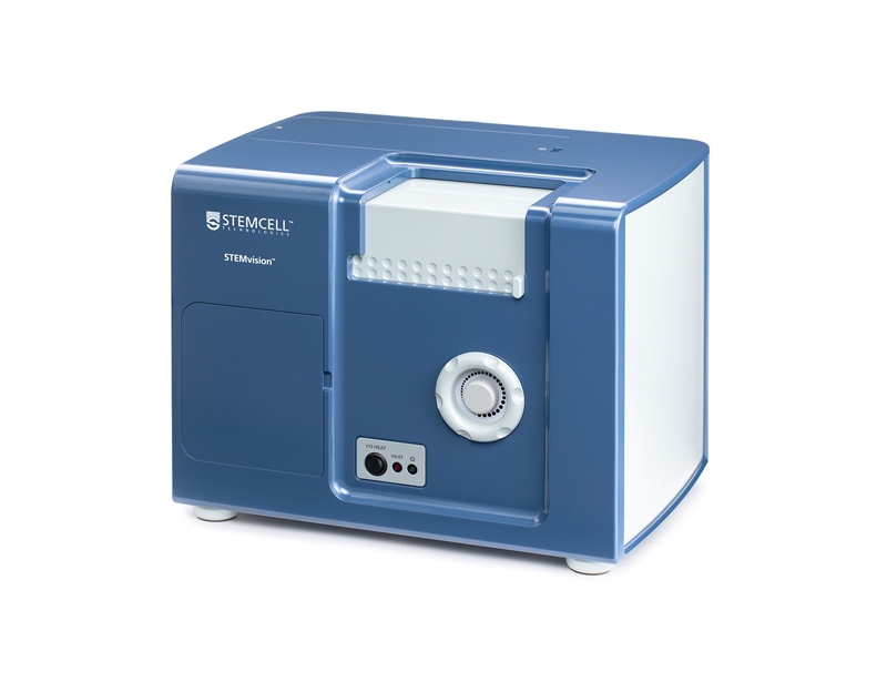
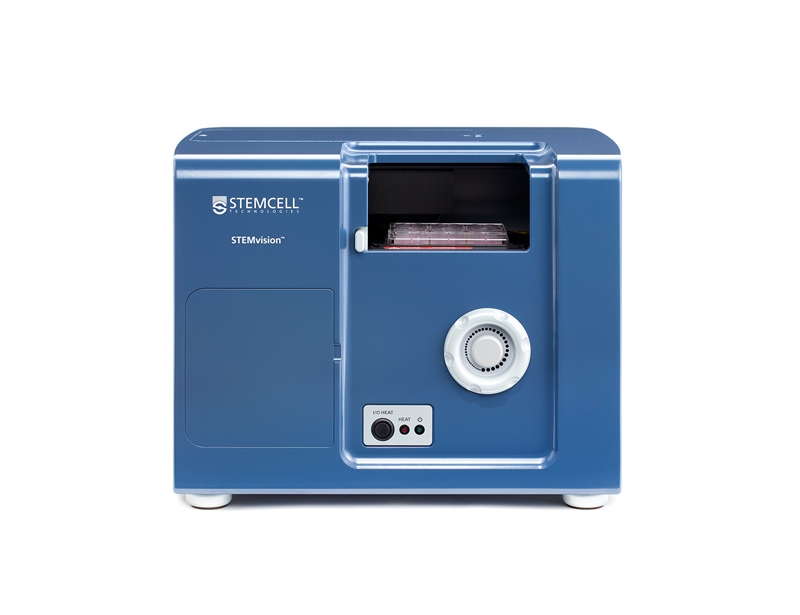
STEMvision™

Automated and standardized colony counting for the hematopoietic colony-forming unit (CFU) assay
概要
STEMvision™ consists of an instrument and software designed specifically for imaging and counting colonies in hematopoietic colony-forming unit (CFU) assays using MethoCult™ media and meniscus-free SmartDish™ cultureware. STEMvision™ software, known as Analysis Packages, replaces the need to manually count colonies using a microscope. With the addition of the STEMvision™ 21 CFR Part 11 Compliance Software Add-On (Catalog #500-0110), STEMvision™ can support workflows consistent with FDA Title 21 CFR Part 11 regulations on electronic records.

For human cultures, three Analysis Packages have been designed for scoring and counting hematopoietic colonies produced by erythroid, myeloid and multi-potential progenitor cells in 14-day assays of CB, BM and MPB cultured in MethoCult™ Optimum media. A fourth Analysis Package is also available for counting the total number of colonies in 7-day assays of CB cultured in MethoCult™ Express medium.

For mouse BM cultures, three Analysis Packages have been designed to count total numbers of hematopoietic colonies produced by all combined myeloid and erythroid progenitors in M3434, myeloid progenitors only in M3534 and erythroid progenitors only in M3436.

Selection of at least one Analysis Package is required for each instrument purchase. Additional Analysis Packages and software add-ons to support 21 CFR Part 11 compliance are sold separately. Select your preferred Analysis Package and select Request Pricing for further information. Several purchase and leasing options are available.

For more information about Instrument Services including additional service packages and installation software please see our instrumentation overview.

Browse our Frequently Asked Questions (FAQs) on performing the CFU assay and explore its utility as part of the cell therapy workflow.                                
Species
Human, Mouse
Application
Colony Assay, Functional Assay, Genome Editing
Brand
STEMvision
Area of Interest
Cell Therapy, Cord Blood Banking, Stem Cell Biology, Transplantation Research
技术资料
Document Type产品名称Catalog #Lot #语言
Manual STEMvision™                                22000AllEnglish
数据及文献

Data

Representative STEMvision™ Images Showing Colonies Derived from CB Progenitors after 7 Days of Culture in MethoCult™ Express, and from CB, BM and MPB after 14 Days of Culture in MethoCult™ Optimum

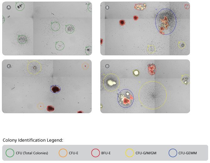
Figure 1. Representative STEMvision™ Images Showing Colonies Derived from CB Progenitors after 7 Days of Culture in MethoCult™ Express, and from CB, BM and MPB after 14 Days of Culture in MethoCult™ Optimum

These images have been analyzed by STEMvision™ Human (A) 7-Day and (B-D) 14-Day Analysis Packages. Green circles identify individual colonies in a 7-day CB CFU assay that scores total CFUs only (A). Orange and red circles identify erythroid colonies, yellow circles identify myeloid colonies and blue circles identify mixed colonies in 14-day CB (B), BM (C) and MPB (D) CFU assays. Erythroid and mixed colonies that contain hemoglobinized cells are shown in true red color.

STEMvision™ Automated Counting of Total, Erythroid (BFU-E) and Myeloid (CFU-G/M/GM) Colonies Is Highly Correlated to Manual Counts of 14-Day CB CFU Assays

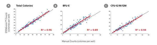
Figure 2. STEMvision™ Automated Counting of Total, Erythroid (BFU-E) and Myeloid (CFU-G/M/GM) Colonies Is Highly Correlated to Manual Counts of 14-Day CB CFU Assays

Cryopreserved CB cells were thawed, plated in MethoCult™ Optimum and cultured for 14 days. The resulting colonies were then counted both manually using an inverted microscope, and automatically using STEMvision™ with the Human CB 14-Day CFU Analysis Package (Catalog #22005). Gray dashed lines represent a perfect linear correlation between manual and automated counts. Red solid lines represent the actual linear correlation between manual and automated counts. The mathematical equations and correlation coefficients (R2) that describe each data set (n=130 CFU assays) are as follows: A: y=1.02x + 1.39, R2=0.96 for Total Colonies, B: y=1.05x + 1.53, R2=0.89 for BFU-E, C: y=0.99x + 0.13, R2=0.94 for CFU-G/M/GM.

STEMvision™ Automated Counting of Mixed Colonies Falls Within the Range of Manual Counts of 14-Day CB CFU Assays

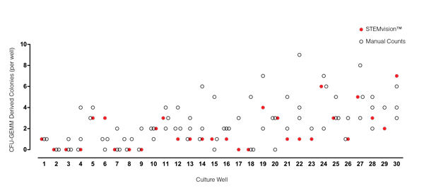
Figure 3. STEMvision™ Automated Counting of Mixed Colonies Falls Within the Range of Manual Counts of 14-Day CB CFU Assays

Thirty individual 14-day CB CFU assays were counted by three to seven people. The numbers of mixed colonies (CFU-GEMM) colonies counted manually in each well are shown by the black open circles (n=80 total assay scores). Manual CFU-GEMM counts in most cultures varied significantly between individual people. STEMvision™ counts of the same cultures (red circles) provided a CFU-GEMM count that was typically within the range of manual counts.

STEMvision™ Automated Scoring of Total, Erythroid (BFU-E + CFU-E) and Myeloid (CFU-G/M/GM) Colonies Is Highly Correlated to Manual Counts of 14-Day BM CFU Assays

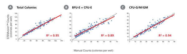
Figure 4. STEMvision™ Automated Scoring of Total, Erythroid (BFU-E + CFU-E) and Myeloid (CFU-G/M/GM) Colonies Is Highly Correlated to Manual Counts of 14-Day BM CFU Assays

Cryopreserved BM cells were thawed, plated in MethoCult™ Optimum, cultured for 14 days, and the resulting colonies then scored both manually using an inverted microscope and automatically using STEMvision™ with the Human BM 14-Day CFU Analysis Package (Catalog #22006). The BM Analysis Package can identify and count erythroid colonies produced by CFU-E and BFU-E separately, but these are combined in panel B. Gray dashed lines represent a perfect linear correlation between manual and automated counts. Red solid lines represent the actual linear correlation between manual and automated counts. The mathematical equations and correlation coefficients (R2) that describe each data set (n=120 CFU assays) are as follows: A: y=0.88x + 8.79, R2=0.95 for Total Colonies, B: y=0.83x + 6.71, R2=0.89 for CFU-E + BFU-E, C: y=0.92x + 2.55, R2=0.94 for CFU-G/M/GM.

STEMvision™ Automated Counting of Mixed Colonies Falls Within the Range of Manual Counts of 14-Day BM CFU Assays

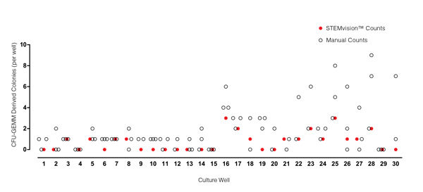
Figure 5. STEMvision™ Automated Counting of Mixed Colonies Falls Within the Range of Manual Counts of 14-Day BM CFU Assays

Thirty individual 14-day BM CFU assays were counted by three to seven people. The numbers of mixed (CFU-GEMM) colonies counted manually in each well is shown by the black open circles (n=82 total assay scores). Manual CFU-GEMM counts in most cultures varied significantly between individual people. STEMvision™ counts of the same cultures (red circles) provided a CFU-GEMM count that was typically within the range of manual counts.

STEMvision™ Automated Counting of Total, Erythroid (BFU-E) and Myeloid (CFU-G/M/GM) Colonies Is Highly Correlated to Manual Counts of 14-Day MPB CFU Assays

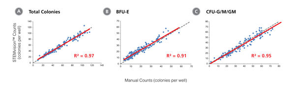
Figure 6. STEMvision™ Automated Counting of Total, Erythroid (BFU-E) and Myeloid (CFU-G/M/GM) Colonies Is Highly Correlated to Manual Counts of 14-Day MPB CFU Assays

Cryopreserved MPB cells were thawed, plated in MethoCult™ Optimum, cultured for 14 days, and the resulting colonies then scored both manually using an inverted microscope and automatically using STEMvision™ with the Human MPB 14-Day CFU Analysis Package (Catalog #22007). Gray dashed lines represent a perfect linear correlation between manual and automated counts. Red solid lines represent the actual linear correlation between manual and automated counts. The mathematical equations and correlation coefficients (R2) that describe each data set (n=143 CFU assays) are as follows: A: y=0.97x + 2.44, R2=0.97 for Total Colonies, B: y=0.96x + 3.74, R2=0.91 for BFU-E, C: y=0.96x + 0.90, R2=0.95 for CFU-G/M/GM.

STEMvision™ Automated Scoring of Mixed Colonies Falls Within the Range of Manual Counts of 14-Day MPB CFU Assays

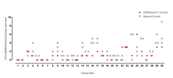
Figure 7. STEMvision™ Automated Scoring of Mixed Colonies Falls Within the Range of Manual Counts of 14-Day MPB CFU Assays

Thirty individual 14-day MPB CFU assays were counted by three to seven people. The numbers of mixed (CFU-GEMM) colonies counted manually in each well is shown by the black open circles (n=82 total assay scores). Manual CFU-GEMM counts in most cultures varied significantly between individual people. STEMvision™ counts of the same cultures (red circles) provided a CFU-GEMM count that was typically within the range of manual counts.

Representative STEMvision™ Images Showing Colonies Derived from Mouse BM Progenitors After 12 Days of Culture in MethoCult™ GF M3434, MethoCult™ GF M3534 or MethoCult™ SF M3436 Media

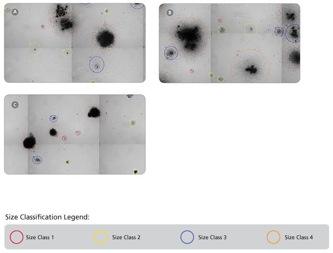
Figure 8. Representative STEMvision™ Images Showing Colonies Derived from Mouse BM Progenitors After 12 Days of Culture in MethoCult™ GF M3434, MethoCult™ GF M3534 or MethoCult™ SF M3436 Media

Images of mouse BM cells cultured in (A) MethoCult™ GF M3434, (B) MethoCult™ GF M3534 and (C) MethoCult™ SF M3436 were acquired using STEMvision™. The corresponding STEMvision™ Mouse Analysis Package (Table 1) was used to analyze each image. Red circles identify the smallest colonies - size class 1, yellow circles - size class 2, blue circles - size class 3 and orange circles identify the largest colonies - size class 4.

STEMvision™ Automated Counting is Highly Correlated to Manual Counting of Total (Myeloid Plus Erythroid) Colonies in Mouse BM CFU Assays

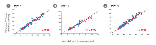
Figure 9. STEMvision™ Automated Counting is Highly Correlated to Manual Counting of Total (Myeloid Plus Erythroid) Colonies in Mouse BM CFU Assays

BM cells were plated in MethoCult™ GF M3434 (Catalog #03434/03444). Colonies were counted on days (A) 7, (B) 10 and (C) 12 both manually using an inverted microscope, and automatically using STEMvision™ equipped with the Mouse Total CFU Analysis Package (Catalog #22008). We recommend counting CFU assays of mouse progenitor cells plated in M3434 between 10 and 12 days. Gray dashed lines represent a theoretical perfect linear correlation between manual and automated counts. Red solid lines represent the actual linear correlation between manual and automated counts. The slope, 95% confidence intervals, correlation coefficients (R2) and sample size for each data set are provided in Table 1.

STEMvision™ Automated Counting is Highly Correlated to Manual Counting of Myeloid Colonies in Mouse BM CFU Assays

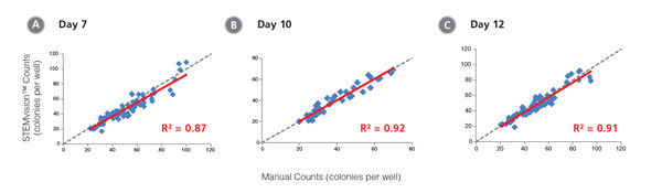
Figure 10. STEMvision™ Automated Counting is Highly Correlated to Manual Counting of Myeloid Colonies in Mouse BM CFU Assays

BM cells were plated in MethoCult™ GF M3534 (Catalog #03534). Colonies were counted on days (A) 7, (B) 10 and (C) 12 both manually using an inverted microscope, and automatically using STEMvision™ equipped with the Mouse Myeloid CFU Analysis Package (Catalog #22009). We recommend counting CFU assays of mouse myeloid progenitor cells plated in M3534 between 10 and 12 days. Gray dashed lines represent a theoretical perfect linear correlation between manual and automated counts. Red solid lines represent the actual linear correlation between manual and automated counts. The slope, 95% confidence intervals, correlation coefficients (R2) and sample size for each data set are provided in Table 1.

STEMvision™ Automated Counting is Highly Correlated to Manual Counting of Erythroid Colonies in Mouse BM CFU Assays

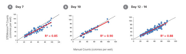
Figure 11. STEMvision™ Automated Counting is Highly Correlated to Manual Counting of Erythroid Colonies in Mouse BM CFU Assays

BM cells were plated in MethoCult™ SF M3436 (Catalog #03436). Colonies were counted on days (A) 7, (B) 10 and (C) 12 - 14 both manually using an inverted microscope, and automatically using STEMvision™ equipped with the Mouse Erythroid CFU Analysis Package (Catalog #22011). We recommend counting CFU assays of mouse erythroid progenitor cells plated in M3436 between of 10 to 14 days. Gray dashed lines represent a theoretical perfect linear correlation between manual and automated counts. Red solid lines represent the actual linear correlation between manual and automated counts. The slope, 95% confidence intervals, correlation coefficients (R2) and sample size for each data set are provided in Table 1.

Table 1. Correlation Between Automated STEMvision™ and Manual Colony Counting

Table 1. Correlation Between Automated STEMvision™ and Manual Colony Counting

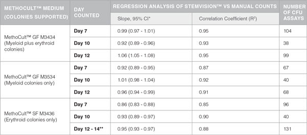
*CI: Confidence Internal
**Mouse CFU assays of erythroid progenitor cells plated in M3436 should be counted between 10 to 14 days.

Publications (6)

Cell stem cell                                        2019                                        mar                                    The NAD-Booster Nicotinamide Riboside Potently Stimulates Hematopoiesis through Increased Mitochondrial Clearance.                                    N. Vannini et al.                                    

Abstract

It has been recently shown that increased oxidative phosphorylation, as reflected by increased mitochondrial activity, together with impairment of the mitochondrial stress response, can severely compromise hematopoietic stem cell (HSC) regeneration. Here we show that the NAD+-boosting agent nicotinamide riboside (NR) reduces mitochondrial activity within HSCs through increased mitochondrial clearance, leading to increased asymmetric HSC divisions. NR dietary supplementation results in a significantly enlarged pool of progenitors, without concurrent HSC exhaustion, improves survival by 80{\%}, and accelerates blood recovery after murine lethal irradiation and limiting-HSC transplantation. In immune-deficient mice, NR increased the production of human leucocytes from hCD34+ progenitors. Our work demonstrates for the first time a positive effect of NAD+-boosting strategies on the most primitive blood stem cells, establishing a link between HSC mitochondrial stress, mitophagy, and stem-cell fate decision, and unveiling the potential of NR to improve recovery of patients suffering from hematological failure including post chemo- and radiotherapy.
Cytotherapy                                        2019                                        jun                                    Validation of a semi automatic device to standardize quantification of Colony-Forming Unit (CFU) on hematopoietic stem cell products.                                    M. Velier et al.                                    

Abstract

Accurate characterization of hematopoietic stem cells (HSC) products is needed to better anticipate the hematopoietic reconstitution and the outcome in patients. Although CD34+ viable cells enumeration is a key predictor of time to correction of aplasia, it does not fully inform about functionality of cells contained in the graft. CFU assay is the gold standard in vitro potency assay to assess clonogenicity of HSC and consists on the count and identification of colonies several days after culture in a semi solid media. Manual count of colonies with optic microscope is the most commonly used method but its important variability and subjectivity hinders the universal implementation of this potency assay. The aim of this study is to validate a standardized method using the STEMvision™ system, the first semi-automated instrument for imaging and scoring hematopoietic colonies, according to French and European recommendations. Results obtained highlight better performance criteria with STEMvision™ system than the manual method. This semi-automatic device tends to reduce the coefficients of variation of repeatability, inter-operator variability and intermediate precision. This newly available platform could represent an interesting option, significantly improving performances of CFU assays used for the characterization of hematopoietic progenitors.
Scientific reports                                        2018                                        NOV                                    TRIAMF: A New Method for Delivery of Cas9 Ribonucleoprotein Complex to Human Hematopoietic Stem Cells.                                    J. Yen et al.                                    

Abstract

CRISPR/Cas9 mediated gene editing of patient-derived hematopoietic stem and progenitor cells (HSPCs) ex vivo followed by autologous transplantation of the edited HSPCs back to the patient can provide a potential cure for monogenic blood disorders such as $\beta$-hemoglobinopathies. One challenge for this strategy is efficient delivery of the ribonucleoprotein (RNP) complex, consisting of purified Cas9 protein and guide RNA, into HSPCs. Because $\beta$-hemoglobinopathies are most prevalent in developing countries, it is desirable to have a reliable, efficient, easy-to-use and cost effective delivery method. With this goal in mind, we developed TRansmembrane Internalization Assisted by Membrane Filtration (TRIAMF), a new method to quickly and effectively deliver RNPs into HSPCs by passing a RNP and cell mixture through a filter membrane. We achieved robust gene editing in HSPCs using TRIAMF and demonstrated that the multilineage colony forming capacities and the competence for engraftment in immunocompromised mice of HSPCs were preserved post TRIAMF treatment. TRIAMF is a custom designed system using inexpensive components and has the capacity to process HSPCs at clinical scale.
Nature communications                                        2018                                                                            Myelo-lymphoid lineage restriction occurs in the human haematopoietic stem cell compartment before lymphoid-primed multipotent progenitors.                                    S. Belluschi et al.                                    

Abstract

Capturing where and how multipotency is lost is crucial to understand how blood formation is controlled. Blood lineage specification is currently thought to occur downstream of multipotent haematopoietic stem cells (HSC). Here we show that, in human, the first lineage restriction events occur within the CD19-CD34+CD38-CD45RA-CD49f+CD90+ (49f+) HSC compartment to generate myelo-lymphoid committed cells with no erythroid differentiation capacity. At single-cell resolution, we observe a continuous but polarised organisation of the 49f+ compartment, where transcriptional programmes and lineage potential progressively change along a gradient of opposing cell surface expression of CLEC9A and CD34. CLEC9AhiCD34lo cells contain long-term repopulating multipotent HSCs with slow quiescence exit kinetics, whereas CLEC9AloCD34hi cells are restricted to myelo-lymphoid differentiation and display infrequent but durable repopulation capacity. We thus propose that human HSCs gradually transition to a discrete lymphoid-primed state, distinct from lymphoid-primed multipotent progenitors, representing the earliest entry point into lymphoid commitment.
Frontiers in bioengineering and biotechnology                                        2016                                                                            An Efficient Electroporation Protocol for the Genetic Modification of Mammalian Cells.                                    L. Chicaybam et al.                                    

Abstract

Genetic modification of cell lines and primary cells is an expensive and cumbersome approach, often involving the use of viral vectors. Electroporation using square-wave generating devices, like Lonza's Nucleofector, is a widely used option, but the costs associated with the acquisition of electroporation kits and the transient transgene expression might hamper the utility of this methodology. In the present work, we show that our in-house developed buffers, termed Chicabuffers, can be efficiently used to electroporate cell lines and primary cells from murine and human origin. Using the Nucleofector II device, we electroporated 14 different cell lines and also primary cells, like mesenchymal stem cells and cord blood CD34+, providing optimized protocols for each of them. Moreover, when combined with sleeping beauty-based transposon system, long-term transgene expression could be achieved in all types of cells tested. Transgene expression was stable and did not interfere with CD34+ differentiation to committed progenitors. We also show that these buffers can be used in CRISPR-mediated editing of PDCD1 gene locus in 293T and human peripheral blood mononuclear cells. The optimized protocols reported in this study provide a suitable and cost-effective platform for the genetic modification of cells, facilitating the widespread adoption of this technology.
The Journal of experimental medicine                                        2014                                        AUG                                    RHEX, a novel regulator of human erythroid progenitor cell expansion and erythroblast development.                                    Verma R et al.                                    

Abstract

Ligation of erythropoietin (EPO) receptor (EPOR) JAK2 kinase complexes propagates signals within erythroid progenitor cells (EPCs) that are essential for red blood cell production. To reveal hypothesized novel EPOR/JAK2 targets, a phosphotyrosine (PY) phosphoproteomics approach was applied. Beyond known signal transduction factors, 32 new targets of EPO-modulated tyrosine phosphorylation were defined. Molecular adaptors comprised one major set including growth factor receptor-bound protein 2 (GRB2)-associated binding proteins 1-3 (GAB1-3), insulin receptor substrate 2 (IRS2), docking protein 1 (DOK1), Src homology 2 domain containing transforming protein 1 (SHC1), and sprouty homologue 1 (SPRY1) as validating targets, and SPRY2, SH2 domain containing 2A (SH2D2A), and signal transducing adaptor molecule 2 (STAM2) as novel candidate adaptors together with an ORF factor designated as regulator of human erythroid cell expansion (RHEX). RHEX is well conserved in Homo sapiens and primates but absent from mouse, rat, and lower vertebrate genomes. Among tissues and lineages, RHEX was elevated in EPCs, occurred as a plasma membrane protein, was rapidly PY-phosphorylated textgreater20-fold upon EPO exposure, and coimmunoprecipitated with the EPOR. In UT7epo cells, knockdown of RHEX inhibited EPO-dependent growth. This was associated with extracellular signal-regulated kinase 1,2 (ERK1,2) modulation, and RHEX coupling to GRB2. In primary human EPCs, shRNA knockdown studies confirmed RHEX regulation of erythroid progenitor expansion and further revealed roles in promoting the formation of hemoglobinizing erythroblasts. RHEX therefore comprises a new EPO/EPOR
Top